Image: Anna Jurkovska/Shutterstock
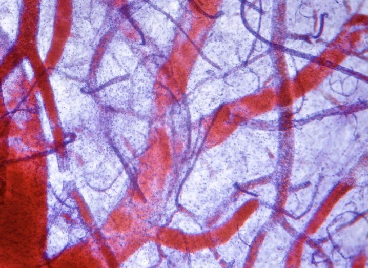
Researchers in Austria have made a medical breakthrough, with new techniques to visualise blood vessels in detail speeding up diagnostic capabilities.
Getting 3D imagery of blood vessels has proved elusive to medical scientists, thanks to the windy route that technology must take to accurately map a moveable feast.
However researchers may now have solved the problem, thanks to a novel approach developed by Miloš Šrámek, who is based at the Austrian Academy of Sciences.
Through new algorithms developed by Šrámek and his team, continuous imagery of blood-vessel lumen is possible. This could therefore help doctors hone in on where blockages exist far faster than they can now.
The two processes developed to help map vessels are called centerline reformation and curved surface reformation.
Through the former, new algorithms of image-processing enable the continuous representation of the interior of a blood vessel.
“Using this technique, we are actually able to visualise the lumen or inside of lung and brain blood vessels without overlaps – an approach that was considered extremely difficult up to now, due to the sharp twists and turns,” said Rüdiger Schernthaner from the Medical University of Vienna.
Curved surface reformation then helps form the 3D visualisation, through a process called ‘ray casting’, which enables the rapid visualisation of 3D objects.
Eduard Gröller of Technische Universität Wien, who also worked on the project, said visualisations with “low-level distortion” are possible, thanks to the algorithms establishing the dimensions of “adjacent anatomical structures”.
“This allows the doctor to identify extravascular pathologies while examining the blood vessels,” he said, while noting that the adoption of appropriate “levels of detail”, which process information near the centerline of a blood vessel with high accuracy, was crucial to this development.
Diagnostic tools are improving at a really high rate in recent years and months, with remote monitoring, microfish swimming through your veins and sensory implants being the focus of detailed attention from medical professionals all over the world.
Though allowing doctors to see 3D renders of patients’ blood vessels, rather than trawling through wave after wave of CT images, could be the best development yet.