Image: © nobeastsofierce/Stock.adobe.com
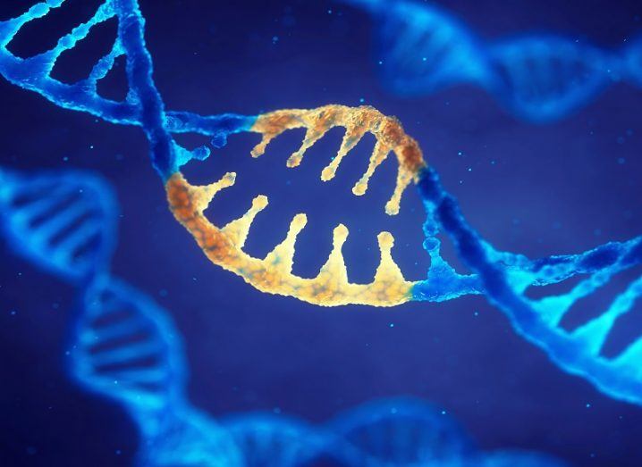
For the first time, the before and after images of the Cas9 enzyme used in CRISPR gene editing have been captured in clear detail.
As a technology, CRISPR remains a big unknown for geneticists who are only now just scratching the surface of what it could achieve. The gene editing tool – which uses an enzyme called Cas9 to precisely cut and edit sequences of DNA – made headlines last year after a Chinese scientist admitted to using it on unborn children to make them resistant to HIV.
The wider scientific community reacted with outrage at the time, warning that there could be many unintended genetic consequences to altering human DNA using the tool. However, a team of scientists from the University of Illinois at Chicago has revealed a world first that could help drastically improve CRISPR’s efficiency and accuracy.
In a paper published to Nature Structural and Molecular Biology, the team said it has captured atomic-level 3D images of Cas9 before and after cutting the DNA. These images provide new structural information on how the enzyme works and gives researchers a much greater understanding of what they’re working with.
‘We have a much clearer picture’
“One of the main hurdles preventing the development of better gene-editing tools using Cas9 is that we didn’t have any images of the enzyme after the cutting DNA and did not have complete information about the changes this very important enzyme undergoes to execute the reaction,” said Miljan Simonovic, a corresponding author of the study.
Previous structural images of Cas9 were obtained using x-ray crystallography which comes with a number of limitations. For example, to capture various states of the enzyme in crystalline form, researchers would have had to use either inactive Cas9 or form the crystals under conditions that don’t support DNA development. In other words, these images only provide a snapshot of before, but not after, the cut.
For this research, the team used cryogenic electron microscopy known for its ability to image large molecules at high-resolution. Activated Cas9 complexes were then flash frozen and captured as images. Most remarkably, in two of the three states, the target DNA was cleaved but the enzyme remained bound to it, allowing this final step of the biochemical reaction sequence to be observed with high resolution.
“Researchers interested in either modulating Cas9 activity or engineering mutant enzymes that might work better just didn’t have complete information to begin with, so advancement was not as fast as desired,” Simonovic said. “But now we have a much clearer picture.”