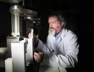
Marios Georgiadis, PhD student at ETH Zurich, Institute for Biomechanics, takes a closer look at a silicon wafer containing dozens of microfluidic probes. Photo by IBM Research - Zurich
A flexible, non-contact microfluidic probe made from silicon that can help researchers and pathologists examine tissue samples is aiming to minimise patient discomfort and provide more accurate results.
Developed by IBM scientists, the microfluidic probe can stain tissue sections at the micrometer scale, performed for drug discovery and disease diagnostics.
“This capability allows the clinician to not only do more with a smaller sample, but will also allow the use of multiple stains on the same sample, therefore increasing the accuracy of the diagnosis,” said Prof Dr Ali Khademhosseini, associate professor at Harvard Medical School and Brigham and Women’s Hospital.
“Thus this work may be transformative for diagnosing a variety of ailments ranging from cancer to cardiac disease.”
Tissue staining is widely used in pathology to detect disease markers in a patient’s sample. More specifically, a particular disease marker is bound with an antibody, which is then chemically coloured or stained on the tissue, IBM said. The colour’s intensity classifies and determines the extent of a disease.
However, tissue staining can be a tedious process, involving many chemical steps, and obtaining a biopsy is an invasive procedure for the patient, so small samples are taken whenever possible.
Enter the microfluidic probe that IBM scientists in Zurich have reported on in the peer-reviewed journal Lab on a Chip.
Measuring 8 millimetres, the diamond-shaped probe consists of a silicon microfluidic head with two microchannels at each tip. The head injects the liquid on the sample’s surface, and continuously aspirates the liquid to prevent it spreading and accumulating on the sample, which can lead to overexposure, IBM said.
Specifically for tissue section analysis, the probe can deliver an antibody very locally in a selected area of a tissue section. Since analysis can be done on spots and lines instead of on the entire tissue section, the tissue is better preserved for additional tests, if required. In addition, only a few picoliters (one trillionth of a litre) of liquid containing antibodies are needed for each analysis spot.
IBM scientists will continue to test and improve the probe and potentially begin using it in laboratory environments in the next few months. The team also plans to explore specific clinical applications, possibly with partners in the field of pathology.
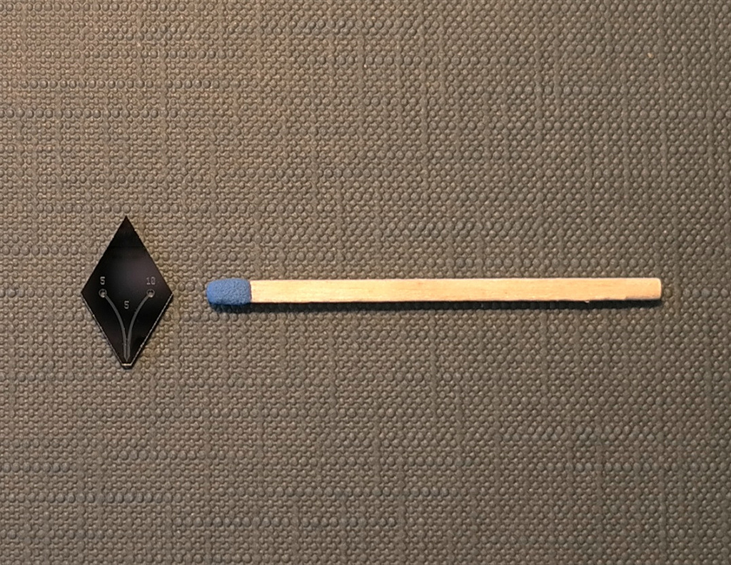
The microfluidic probe consists of a silicon microfluidic head having two microchannels. The head re-aspirates the liquid that it injects on a surface. This prevents spreading and accumulation of the liquid on the surface, which can lead to overexposure. Photo by IBM Research – Zurich