Chronic traumatic encephalopathy (CTE) is a neurodegenerative condition that is attracting immense attention of late. Now, thanks to researchers from Trinity College Dublin (TCD), attempts to diagnose it could improve dramatically.
We recently looked at brain injuries following careers in impact sport, on the back of mainstream interest in a part of health that was previously underappreciated.
Amid a growing number of former American Football players claiming to have been left with neurological conditions due to impacts suffered while playing the sport – with sports like rugby, boxing and mixed martial also seeing issues being raised – scientists are battling to accurately diagnose these types of issues.
Not scientific enough
The problem, Prof Matthew Campbell of TCD told me, is there is no diagnostic criteria to diagnose CTE while the patient is alive.
“No, we’re in a situation where doctors meet patients who tell them of bangs on the head they had 10 years ago, which may lead to CTE,” he said. “It’s not scientific enough.”
Largely speaking, at the moment, tiny slivers of brains are tested, post-mortem, to determine CTE. However that could all change soon, with Campbell’s paper in the Journal of Neuropathology & Experimental Neurology highlighting a potential new route to diagnosis, which can be done “in life”.
“To diagnose CTE, doctors look for a molecule in the brain called TAU,” he said, describing it as like the elastic band that holds asparagus together in a store. “Yes, it holds together microtubules like that.”
Evidence of this leads to evidence of brain injury, which helps determine CTE on a microscopic, post-mortem basis.
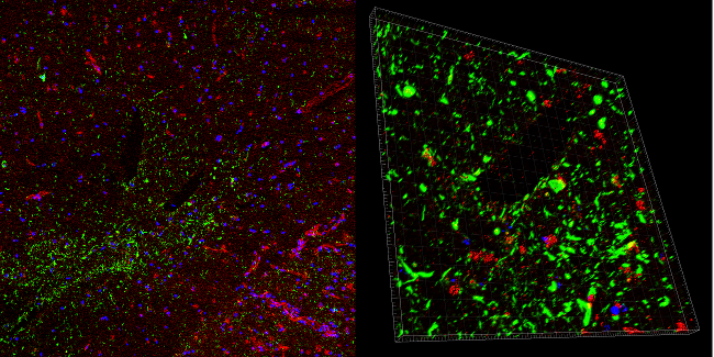
Left: Characteristic staining of p-Tau (green) in a case of CTE. Claudin-5 (red) protein levels are discontinuous and not present in areas of p-Tau accumulation around blood vessels. This is an indication of a dysfunctional blood brain barrier. Right: Areas of p-Tau (green) staining around blood vessels have absent or discontinuous claudin-5 (red) staining indicating a compromised blood brain barrier.
The brain is special
An important thing to note is blood vessels in the brain are unlike vessels in other areas of the body, having unique properties that tightly regulate what gets in or out of the delicate neural tissue.
This is termed the blood-brain barrier (BBB), and a compromised BBB has been linked to numerous other neurological disorders, such as Alzheimer’s disease and multiple sclerosis (MS).
According to this new piece of research, Campbell and his colleagues discovered TAU built up around blood vessels of a brain, which they investigated post-mortem. What’s key about this is it gives a localised area for doctors to search for in living patients.
Compromised BBBs can, if found, highlight possible CTE, should the paper by Campbell and his colleagues prove conclusive.
“We have contrast enhancement MRI machines. With this, it gives us the clinical tools in life to help diagnose,” said Campbell.
This is by no means a definitive green light that doctors can use, merely a case report. However, it’s a flashpoint, something which further research may help confirm.
Prof Michael Farrell, consultant neuropathologist and co-director of the Dublin Brain Bank, said: “Understanding how CTE develops will be critical to identifying new ways to detect its onset in living subjects in addition to developing therapies for this rare but socially important brain disorder.”
Brain injury image via Shutterstock