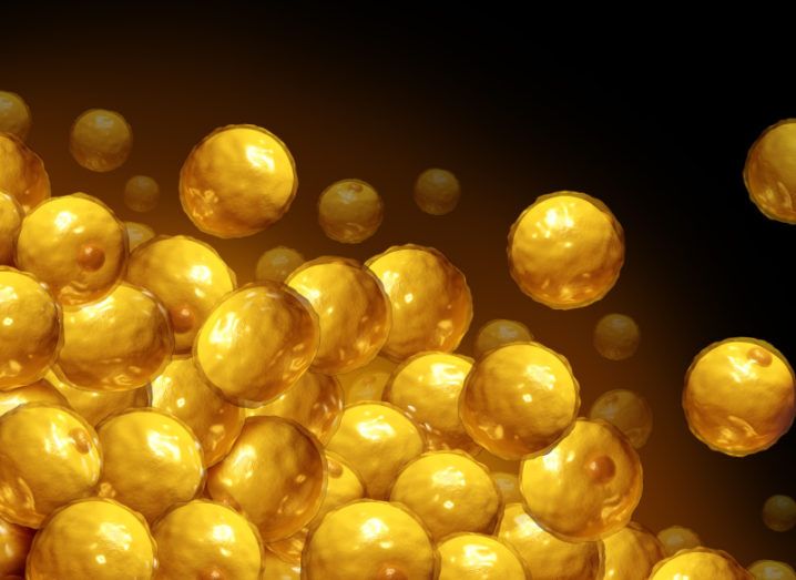
A cluster of fat cells. Image: © freshidea/Stock.adobe.com
Researchers said their work was ‘truly venturing into the unknown’ as they combined immunology and neuroscience to examine visceral fat.
Fat has a tricky reputation. While some fat cells are needed to sustain us, visceral fat can be particularly problematic. This envelops our vital organs but, if is too abundant, produces unhealthy levels of proteins and hormones that negatively affect the tissues and organs around it.
Visceral fat stores have been linked to different forms of cancer, including breast and colorectal, as well as cardiovascular disease.
But understanding the biological processes underpinning visceral fat is no easy task. This was the goal of new research published yesterday (18 August) in the journal Nature, where scientists used mice to study the neuroimmune process involved in the body’s stores of visceral fat.
“Excess visceral fat is very dangerous and at the same time very difficult to eliminate,” explained Henrique Veiga-Fernandes, principal investigator and co-director at the Champalimaud Research Programme in Portugal.
“In this project, our team set out to explore the mechanisms that naturally reduce it, with the hope of uncovering potential clinical applications.”
Back to basics
Understanding the structure of visceral fat tissue was the first step in the research. The tissue is comprised of fat cells, immune cells, nerve fibres and a variety of other cells. One immune type cell of particular interest for the team was Type 2 Innate Lymphoid Cells (ILC2s).
“ILC2s are essential for various immune functions in many tissues and organs, including maintaining the overall wellbeing of fat tissue. However, we didn’t know which cells control ILC2s in visceral fat and what molecular messages they use to communicate,” explained Ana Filipa Cardoso, the first author of the study.
The lab previously found that in the lungs, it was the nervous system that was responsible for controlling the activity of ILC2s. Much to the team’s surprise, this wasn’t the case in visceral fat. In fat, there was no direct communication between neurons and the immune cells.
What they found was that Mesenchymal cells (MSCs) were acting as a mediator between these immune cells and the neurons.
“Mesenchymal cells have been widely ignored until about one to two decades ago,” said Veiga-Fernandes. “The widespread view was that they mainly produced the scaffolding of the tissue, over which other cells would ‘do the work’. However, scientists have since discovered that MSCs carry out multiple essential active roles.”
Understanding the sequence
Through experimentation, the researchers discovered the cascade of events within visceral fat. First, the neural signals communicate to the MSCs. These MSCs then send the message onto the ILC2s, which in turn tells the fat cells to increase their fat metabolism.
With these mechanisms understood, the team was ready to look at the broader picture and try figure out what was causing the neural activity to store visceral fat.
The team knew that the nerve fibres within visceral fat are controlled by the peripheral nervous system, which is in charge of processes such as regulating blood pressure. This system is driven by the central nervous system. This was why the researchers looked to the brain as the next step of the puzzle.
‘The most challenging thing in a project like this one is that you’re truly working at the frontier. This is not immunology anymore, and it’s not neuroscience either’
–DR HENRIQUE VEIGA-FERNANDES
There, they isolated the paraventricular nucleus in the hypothalamus (PVH) as the culprit. The hypothalamus is known for its wide range of functions, from metabolism and reproduction all the way to cardiovascular functions.
Putting the science in context
“This finding is quite significant. It’s the first clear example of a cross-body neuronal circuit that translates brain information into an obesity-related immune function. It also raises many new questions,” said Veiga-Fernandes.
“For instance, what triggers the PVH to issue the ‘fat burning’ command? Is it something related to behaviour, such as eating certain foods or exercising? Or is it dependent on internal metabolic signals? Or both? It’s a white canvas – we don’t know what it is, and it’s tremendously fascinating.”
The team highlighted the potential applications that could arise from understanding these mechanisms, as it creates multiple access points into the processes that cause visceral fat.
“The most challenging thing in a project like this one is that you’re truly working at the frontier. This is not immunology anymore, and it’s not neuroscience either. You have to master technology, methods and approaches that are cross-disciplinary or multidisciplinary,” Veiga-Fernandes said.
“Some of them don’t even exist, and you have to develop them by scratch. Yet, at the same time, the conceptual challenge is exhilarating; we are truly venturing into the unknown.”