Robber fly in macroscopy, at 30x. Image: Jan Rosenboom/Nikon Small World Photomicrography Competition
This year’s Nikon Small World Photomicrography Competition is soon to announce its 2016 winners, so how about a look through some of the eye-catching candidates?
Nikon’s ‘small world’ is a photography competition like no other, taking the microscopic and plastering it on the main stage.
Open to those interested in microscopy and photography, it brings images with obscure lead characters together with scale – sometimes small, often smaller still.

Founded in 1972, the long-running competition has historically thrown up some outstanding images, with a selection of this year’s finalists showcased below.
Judging was completed during the summer, with a panel including Rachel Link, producer of National Geographic, and Dr Joe Hanson, science writer and science show host in the US.
“This judging panel perfectly reflects the spirit and growth of the competition,” said Eric Flem, communications manager at Nikon Instruments.
“As the competition has evolved with both scientific research and technology advancements throughout the decades, we are excited to have a panel that can speak to the scientific integrity, artistic quality, and photo and video production techniques that each entry encompasses.”
This year’s event will see winners announced tomorrow (19 October), with the organisers’ Instagram page revealing all at 4.00pm IST.
This year, there’s an additional competition to find out what the public likes best, lasting a little longer. You can vote for your favourite image here.
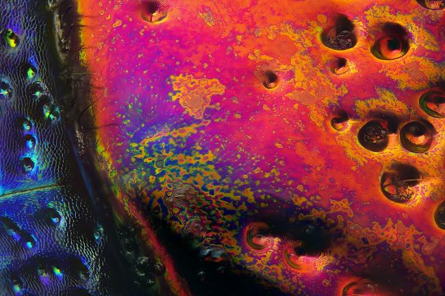
Section of a red speckled jewel beetle (Chrysochroa buqueti rugicollis), in reflected light at 19x. Image: Yousef Al Habshi/Nikon Small World Photomicrography Competition
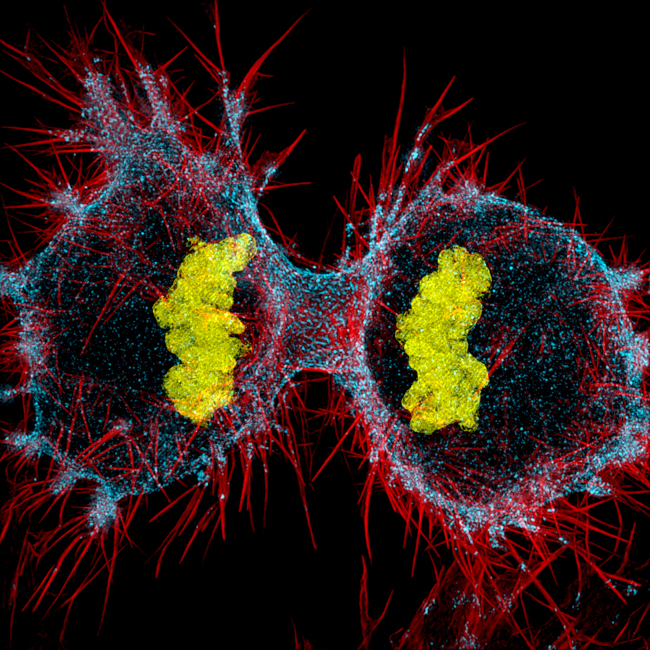
Human HeLa cell undergoing cell division (cytokinesis). DNA (yellow), myosin II (blue) and actin filaments (red), in structured illumination at 9x. Image: Dr Dylan Burnette/Nikon Small World Photomicrography Competition

Ant pupae (Myrmica sp.) in reflected light/focus stacking at 5x. Image: Geir Drange/Nikon Small World Photomicrography Competition
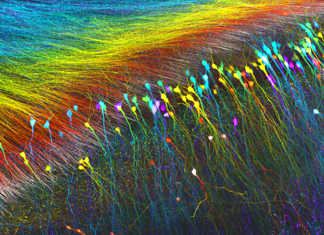
Hippocampal neurons, in confocal at 10x. Image: Dr Wutian Wu/Nikon Small World Photomicrography Competition
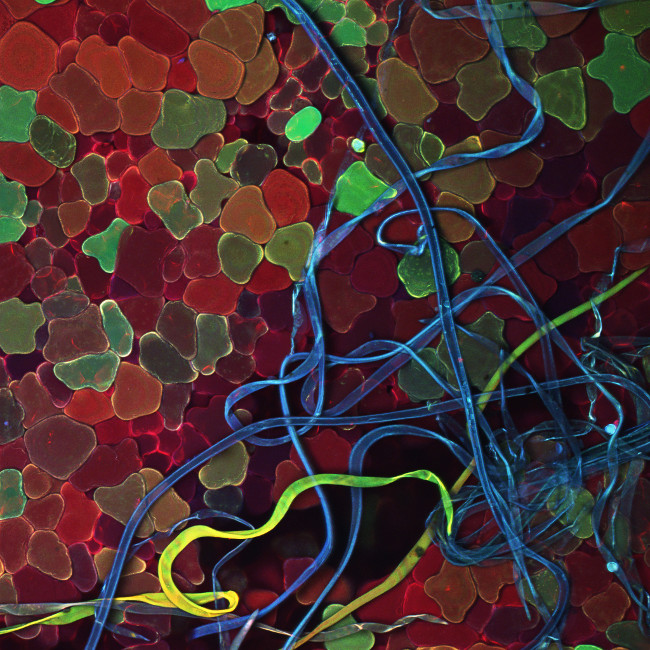
Galls of a mite (Aceria pyracanthi) and fungus on the surface of a scarlet firethorn plant (Pyracantha coccinea) in confocal/fluorescence at 10x. Image: Györgyi Zséli/Nikon Small World Photomicrography Competition
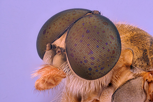
Robber fly in macroscopy, at 30x. Image: Jan Rosenboom/Nikon Small World Photomicrography Competition
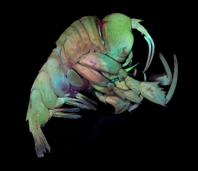
Deep sea crustacea (Phrosina semilunata) in confocal at 40x. Image: Dr Tomonari Kaji/Nikon Small World Photomicrography Competition
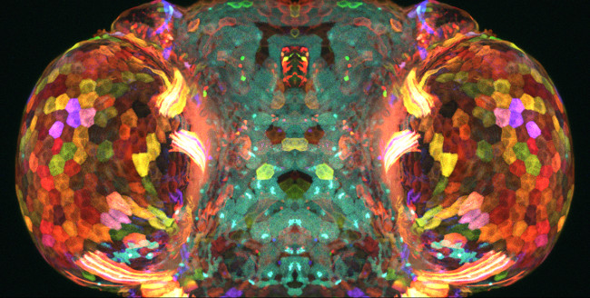
Head of a “skinbow” zebrafish larvae, in confocal at 25x. Image: Dr Chen Chen-Hui/Academia Sinica, Institute of Cellular and Organismic Biology/Nikon Small World Photomicrography Competition

Water mite (Unionicola sp.) in fluroescence at 100x. Image: Jacek Myslowski/Nikon Small World Photomicrography Competition

Green bottle fly, in image stacking at 10x. Image: Erno Endre Gergley/Nikon Small World Photomicrography Competition
If microscopy is not your thing, there’s always the Comedy Wildlife Photo Awards, for which we previously looked through the early candidates.
With more than 1,500 entries last year, the project is a novel way to promote animal conservation throughout the globe. Julian Rad’s ‘Rush Hour’ image of a hamster on the run took last year’s prize in the inaugural edition of the now annual event.