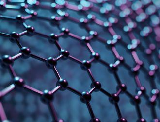
Using ‘spark plasma’ to create 3D solids of graphene could pave the way for vastly improved bone implants in future, according to an international team of scientists.
Researchers based in the US, Brazil and India have banded together to add another string to graphene’s bow, creating 3D structures which, in theory, could replace titanium-based bone implants in future.
They used spark plasma sintering to connect flakes of graphene oxide into porous solids, with the technique utilising a high pulse current – rather than high pressure or temperatures – to weld the material together.

The right fit
The material they made is nearly 50pc porous, with a density half that of graphite and a quarter of titanium metal. Armed with 40 megapascals, however, it has enough ‘compressive strength’ to qualify it for bone implants, said Sekhar Tiwary, co-lead author of a paper published in Advanced Materials describing the technique.
“We started thinking about this for bone implants because graphene is one of the most intriguing materials with many possibilities and it’s generally biocompatible,” he said.
“Four things are important: its mechanical properties, density, porosity and biocompatibility,” he said – areas in which graphene and the spark plasma process excel.
“The nice thing about two-dimensional materials is that they give you a lot of surface area to connect. With graphene, you just need to overcome a small activation barrier to make very strong welds,” he said.
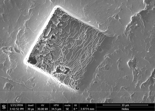
A focused ion beam microscope image shows 3D graphene layers welded together in a block. The material is biocompatible and its material properties meet the standards necessary for consideration as a bone implant, according to researchers at Rice University. Image via Ajayan Group/Rice University
Hard graft
Last week, a team of Irish researchers developed a new way to create bone grafts, 3D bioprinting structures vastly improving on current processes.
The new process uses 3D bioprinting technology to fabricate cartilage templates, which have been shown to assist the growth of a complete bone organ.
The process deposits different biomaterials and adult stem cells in order to engineer cartilage templates, matching the shape of a segment within the build.
Noting the “rapidly expanding” area of 3D bioprinting, AMBER’s Prof Daniel Kelly – following the publication of his paper in Advanced Healthcare Materials – said bone developments have lagged behind those of simple tissues, such as skin, blood vessels and cartilage.
“Our research offers real hope in the future for patients with complex bone trauma or large defects following removal of a tumour.”
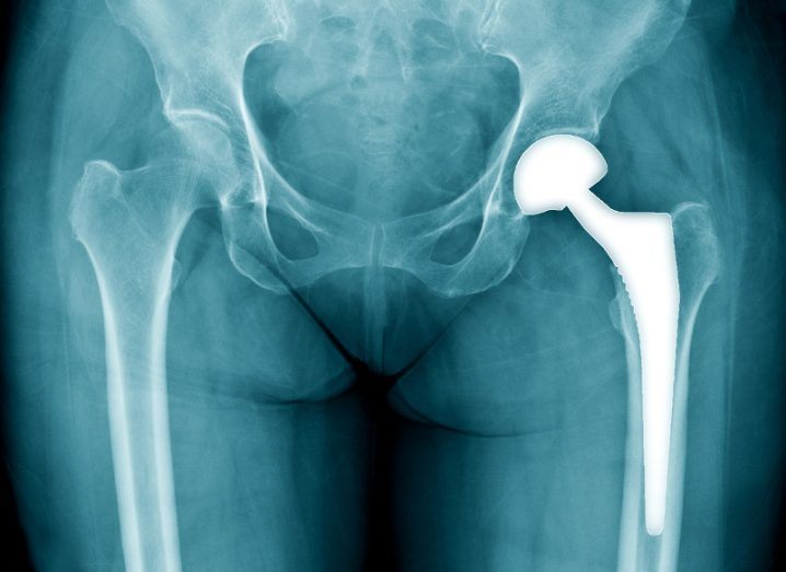
Main hip replacement image via Shutterstock