
Dr Kerry Thompson. Image: Martina Regan Photography
University of Galway’s Dr Kerry Thompson explains how the work of microscopy fits into the wider research community.
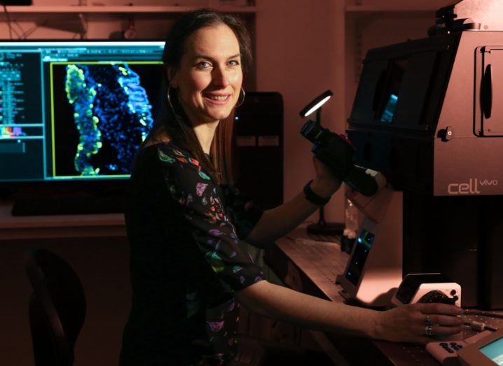
“Microscopy and imaging are fundamental techniques in the global modern research ecosystem,” explains Dr Kerry Thompson, an imaging scientist fellow at the University of Galway. Thompson currently leads development at the Anatomy Imaging and Microscopy (AIM) facility.
AIM is the principal microscopy and imaging research infrastructure in the University of Galway and functions in a dual capacity as both a service provider and centre for research.
In 2020, she was funded by the Chan Zuckerberg Initiative (CZI) to develop training programmes in microscopy.
A large part of her role is bringing the community together “through events at local, national and international levels, looking at the infrastructure landscape and planning strategic acquisitions for the future”.
Thompson was recently awarded €3.6m funding from Science Foundation Ireland and the University of Galway to develop a Centre of Excellence in Multimodal Imaging. As part of the project, three new high-end systems will be brought to campus later this year. “I can’t wait to get the new systems (super resolution, multiphoton deep tissue imaging and volume electron microscopy) up and running.”
‘Images are generally key components of many high-impact publications’
Tell us about your current research.
On a day-to-day basis as a microscopist, I’m usually involved with many multidisciplinary projects at the same time. My engagement with these projects can vary from providing advice on study design to actually capturing the images on the microscope. Every few weeks we get a super interesting new challenge.
At the moment, I’m working closely with my AIM team members on projects for Prof John Dalton and his Molecular Parasitology group, and with Dr Cynthia Coleman on coral containing bioinks for bone regeneration.
Internationally, I’m working with a Global Bioimaging consortium to draft best-practise recommendations for career development and pathways for imaging scientists. We hope to release a paper in the next few months.
In your opinion, why is your research important?
Images are generally key components of many high-impact publications. As professional microscopists, we assist with the collection of temporally and spatially resolved data to place function in a structural context. We provide dynamic and kinetic information that can be correlated with high resolution and high magnification.
The skills, perspectives and training that are offered by core or platform technological scientists underpin much of the research that happens in universities and research institutes. This has really been evidenced by the massive expansion in support for such groupings by philanthropic bodies such as CZI. The pace of growth of technologies has been phenomenal even in the past 10 years.
‘The inherent creativity and freedom of exploration in science really appealed to me’
Three Nobel prizes have been awarded to imaging and microscopy techniques between 2008 and 2017. Along with this, the “quiet revolution” of the volume electron microscopy community, led by researchers such as Dr Lucy Collinson and Prof Paul Verkade, has gained massive pace over a short period of time. Nature has listed it as one of the techniques to watch for 2023.
In Galway, we will bring the first volume electron microscopy techniques to Irish researchers later this year. Coupling that with the advances in bioimage analysis and machine-learning techniques, it’s certainly a very exciting time to be involved in this aspect of microscopy.
What inspired you to become a researcher?
As a child I was fascinated with the natural world. My parents were always incredibly supportive and allowed and encouraged me to indulge my innate (albeit messy) curiosity.
My university experience enabled me to find a field where all the things I love doing collide – science, art and the investigation of tiny things with fascinating structures.
I chose to study biomedical science with a specialisation in human anatomy for my bachelor’s degree. It was an interesting degree, ahead of its time probably, and for which a third of my study was dedicated to information technology.
‘It is true frontier science’
My final year BSc project with Prof Dockery was probably the key turning point for me in realising I wanted to stay in research or academia. The inherent creativity and freedom of exploration in science really appealed to me, along with the beautiful images I realised I could capture to showcase my samples. It was like this huge pick and mix of technologies and techniques to adapt and apply.
I love my work – the more obscure the subject matter the better.
What are some of the biggest challenges or misconceptions you face as a researcher in your field?
Probably one of the biggest challenges we face at the moment is staffing for platforms, along with career development and pathways for this cohort of technological scientists.
There are many ways to become an imaging scientist, and generally no two people have the same story or have taken the same pathway or route to get there. Many are currently employed as technical staff or officers, some as academics, some are more affiliated with administration.
We as a community fit somewhere in between all of these positions. We focus on the development and implementation of new technologies and broadly apply our skills to a huge array of samples and work with multi and transdisciplinary groups. It is true frontier science. This is quite contrary to the expected norm for academics in a traditional university setting. To progress and get promoted, staff often encounter difficulties when benchmarked against normal key performance indicators so we really need to rethink how to support this type of role.
There needs to be sustainable long-term investment in these skilled staff to ensure best use of the demanding technologies we implement and to really ensure our national funders are getting the best possible investment for the national infrastructure.
I’m hoping my work with the Global Bioimaging Careers group will help better inform our policy makers and employers to implement varied and more flexible career structures that take into account the intricacies of this type of role for staff working in this space. Globally, the community will gather in South Africa in October of this year, and we will officially launch this recommendation.
Do you think public engagement with science has changed in recent years?
I think the public have never been more aware of what we do as scientists and researchers or how the scientific method works. Thankfully, my field very much lends itself to public engagement as it is so visual and actually could be considered an art form in itself.
Some of the first images the public saw of the Covid-19 virus were taken on transmission electron microscopes. Microscopy and imaging were fundamental in gaining an understanding of what the virus looked like and how it manipulated cells etc.
I am the honorary secretary for education and outreach with the Royal Microscopical Society (based in Oxford) and we run an international outreach programme for primary school children (in collaboration with the Microscopy Society of Ireland). We lend kits with eight microscopes, activities and samples to schools, free of charge, for a term at a time, and sometimes do school visits, time permitting.
Educators are free to explore their own samples or use the curriculum-aligned activities. Microscopy and imaging ties into so many broader educational themes (literacy, numeracy, descriptive skills and art) other than just the physics, chemistry and biology of science. Through exploring minibeasts or analysing basic onion cells, we hope to capture the imagination of, and inspire, the next generation of microscopists.
I think the public has a right to hear about what taxpayer money is funding and what research teams are working on and likewise, be inspiration points for the next generation. It’s great for kids to see who real scientists and researchers are. This is probably one of the most fun aspects of my job – seeing kids look down a microscope for the first time and focus on something like iridescent butterfly wing scales and get excited. It puts everything else in context and reminds me why I got into this in the first place.
10 things you need to know direct to your inbox every weekday. Sign up for the Daily Brief, Silicon Republic’s digest of essential sci-tech news.


