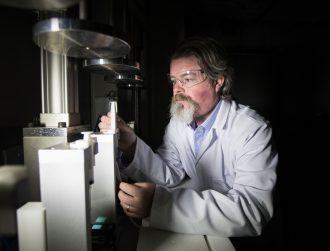
Image: Rui Vieira/PA
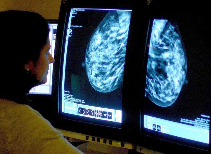
Researchers in the UK have developed a new method of analysing breast tumours in real time to help decide a clearer plan of action.
A scan that involves magnetising molecules can let doctors see in real time which regions of a breast tumour are active, according to new research. Scientists said it is the first time they have shown that this scanning technique – called carbon-13 hyperpolarised imaging – can be used to monitor breast cancer.
It was tested on patients from Addenbrooke’s Hospital in Cambridge with various types and grades of breast cancer before they had received any treatment. Researchers used the scan to measure how fast tumours were metabolising a naturally occurring molecule called pyruvate.
They were able to detect differences in the size, type and grade of tumours – a measure of how fast growing or aggressive the cancer is. The scan was also able to detect variations in metabolism between different regions of the same tumour.
Prof Kevin Brindle, lead researcher from the Cancer Research UK Cambridge Institute, said: “This is one of the most detailed pictures of the metabolism of a patient’s breast cancer that we’ve ever been able to achieve.
“It’s like we can see the tumour ‘breathing’. Combining this with advances in genetic testing, this scan could, in the future, allow doctors to better tailor treatments to each individual and detect whether patients are responding to treatments, like chemotherapy, earlier than is currently possible.”
How it works
Pyruvate is formed in the body from the breakdown of glucose and other sugars. Hyperpolarised carbon-13 pyruvate is a slightly heavier isotope-labelled form of the molecule.
In the study, the scientists hyperpolarised, or magnetised, carbon-13 pyruvate by cooling it to about one degree above absolute zero (minus 273 degrees Celsius) and exposed it to extremely strong magnetic fields and microwave radiation. The frozen material was then thawed and dissolved into an injectable solution.
Patients were injected with the solution and received an MRI scan. Magnetising the carbon-13 pyruvate molecules increases the signal strength by 10,000 times so they are visible on the scan, researchers said.
They used the scan to measure how fast pyruvate was being converted into a substance called lactate. Cells convert pyruvate into lactate as part of the metabolic processes that produce energy and the building blocks for making new cells.
Tumours have a different metabolism to healthy cells and so produce lactate more quickly. This rate also varies between tumours and between different regions of the same tumour.
Heading for clinical trials
According to the study, monitoring this conversion in real-time could be used to infer the type and aggressiveness of the breast cancer.
The team now hopes to trial the scan in larger groups of patients to see if it can be reliably used to inform treatment decisions in hospitals.
Prof Charles Swanton, Cancer Research UK’s chief clinician, said: “This exciting advance in scanning technology could provide new information about the metabolic status of each patient’s tumour upon diagnosis, which could help doctors to identify the best course of treatment.
“And the simple, non-invasive scan could be repeated periodically during treatment, providing an indication of whether the treatment is working. Ultimately, the hope is that scans like this could help doctors decide to switch to a more intensive treatment if needed, or even reduce the treatment dose, sparing people unnecessary side-effects.”
The research, published in the Proceedings of the National Academy of Sciences, was funded by Cancer Research UK.
– PA Media