New imaging software used to detect oesophageal cancer in human samples is being heralded as a “spectacular” success, paving the way for a future 100pc detection rate.
Typically prominent in swathes of Asia and southern Africa, oesophageal cancer is considered one of the most difficult cancers to detect but earlier and earlier detection in recent years has resulted in a slow decline in the number of fatal cases.
However, the software is not perfect and researchers from Eindhoven University of Technology in the Netherlands have been working on a new piece of software that will give medical facilities unprecedented ability to detect the disease early.

Like any disease or cancer, the earlier an abnormality is spotted, the better the chances are of survival and full recovery.
For this particular research, the team looked at how prolonged reflux in patients leads to the development of something called Barrett’s oesophagus which results in abnormal growths that require regular endoscopic hospital checks.
Collaborating with Eindhoven’s Catharina Hospital, the team was able to tinker with its image detection software, which, until that time, had been used for detecting people and objects.
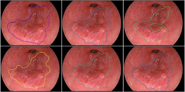
Six contours were drawn by five experts and the computer systems, of early oesophageal cancer. The top-right image is the contour drawn by the computer system. Image via Eindhoven University of Technology
‘Spectacular’ results
Now, with its software tuned to look for the familiar signs of early-stage oesophageal cancer, the initial detection rate has been described as “spectacular” by gastroenterologist, Dr Erik Schoon.
“To recognise early forms of cancer in a Barrett’s oesophagus is one of the most difficult things to do in our field,” he said.
Paving the way for a 100pc detection and treatment rate, the software also saves the patient the strain and dangers of needing surgery to remove part of the oesophagus.
Before it can be rolled out to hospitals, however, the developers and researchers working with the software will need to improve it for analysis real-time video frames of the oesophagus.
It’s envisioned that, once this and other hospital tests are complete, the software will be available for use by medical professionals within the next five to 10 years.
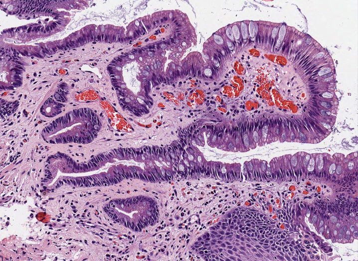
Oesophagus tissue in the microscopic view image via Shutterstock