Image: science photo/Shutterstock
A team of researchers has found a way to use holograms to diagnose chronic diseases remotely, and it’s a lot simpler than you think.
In the near future, patients with chronic diseases with no nearby access to diagnostics could benefit from the remote use of new breakthrough hologram technology and computer science.
The discovery was made by team from the University of California, Los Angeles, which published a paper in the journal Science Advances detailing how – with extremely simple optical hardware, a lens-free microscope and some sophisticated algorithms – accurate reconstructed images of tissue samples can be generated.
This could make badly needed diagnostic testing available and affordable for people in developing countries and remote areas that lack the expensive lab equipment currently used to perform tissue biopsies.
Making the whole process much cheaper
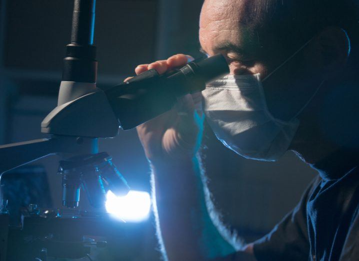
Tissue biopsy is widely considered the gold standard for detecting diseases such as cancer and inflammatory conditions, and involves cutting tissue into thin slices of around one-tenth the thickness of a human hair and detecting abnormalities in cells.
To achieve this, the tissue usually requires fluorescent dyes, which can be costly and can degrade the sample over time.
So, to make this breakthrough, Aydogan Ozcan and his team instead used coloured, light-absorbing dyes that don’t result in the same degradation and can be used with regular microscopy tools.
Then, instead of using a costly biopsy testing machine, the team developed a new device made of components that collectively cost just a few hundred dollars, generating a holographic lens-free microscope that’s capable of producing 3D pictures.
Same tech as used in mobile phones
Even more impressive is that the new method can facilitate tissue samples that can be just 0.2mm thick – 20 times smaller than traditional ones – allowing for a greater sample size.
To actually detect any abnormalities in the samples, the tissue is placed on a silicon chip filled with millions of photo detectors.
When light is shined on the tissue sample, low-resolution shadows fall on the chip, with the results forming holograms of the tissue sample.
These cross-sections can then be analysed in greater detail by researchers to determine any issues.
Yibo Zhang, the study’s first author, said: “Through computation and algorithms, we converted a standard 10-megapixel imager, like those commonly used in mobile phones, into a few-hundred-megapixel microscope that can digitally image through different slices of a thick tissue sample.”