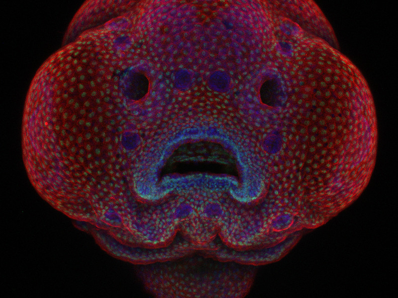

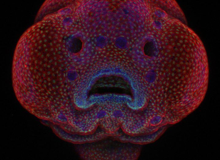
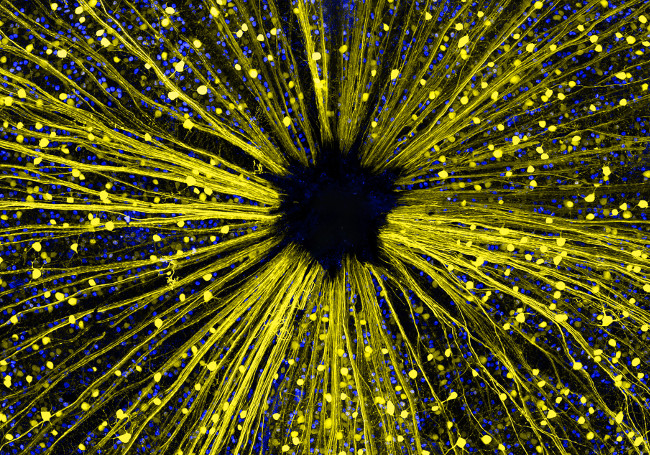
Image: Dr Oscar Ruiz/Nikon Small World Photomicrography Competition
The winner of the Nikon Small World Photomicrography Competition has been revealed, with a zebrafish the star of the show.
Oscar Ruiz took top prize at the 42nd annual Nikon Small World Photomicrography Competition today (19 October), for his microscopic view of the facial development of a four-day-old zebrafish embryo.
Ruiz brings the world face-to-face with his research on facial development and cellular morphogenesis with his image of the zebrafish embryo.

Ruiz used the zebrafish to study genetic mutations that lead to facial abnormalities, such as cleft lip and palate in humans. This research was conducted in the lab of Dr George Eisenhoffer at the University of Texas MD Anderson Cancer Center in Houston, Texas.
The competition is like no other, and aside from Ruiz, the competition recognised 19 other winning images, 15 honourable mentions and 61 images of distinction.
Open to those interested in microscopy and photography, it brings images with obscure lead characters together with scale – sometimes small, often smaller still.
In deciding why Ruiz’s photo should win the top prize, the judges said they were intrigued by his innovative techniques to capture time-lapse images.
Has uses for human patients, too
Using the time-lapse as a guide, Ruiz is creating an atlas of the development of the zebrafish face and with his group, he is tracking physical landmarks throughout to monitor how exactly a normal zebrafish’s face develops.
Similarily, the same metrics can then be used to identify abnormalities in the development of zebrafish harbouring specific genetic mutations identified in human patients.
“Until now, these facial abnormalities had not been extensively studied in a live context where you can see what’s happening during development in real time,” said Ruiz, following his win.
“Using a live-imaging approach means we can better understand and pinpoint exactly how and why these developmental abnormalities occur. The first step is knowing how it happens, then we can figure out how to fix it.”
This year, there’s an additional competition to find out what the public likes best, lasting a little longer. You can vote for your favourite image here.
The 20 best images, in reverse order, are:
20th place – Cow dung in dark field at 30x. Image: Michael Crutchley/ Nikon Small World Photomicrography Competition
19th place – Human neural rosette primordial brain cells, differentiated from embryonic stem cells in confocal at 10x. Image: Dr Gist F Croft, Lauren Pietilla, Stephanie Tse, Dr Szilvia Galgoczi, Maria Fenner, Dr Ali H. Brivanlou/Nikon Small World Photomicrography Competition
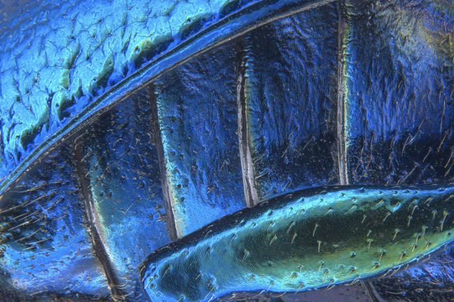
18th place – Parts of wing-cover (elytron), abdominal segments and hind leg of a broad-shouldered leaf beetle (Oreina cacaliae) in stereomicroscopy, image stacking at 40x. Image: Pia Scanlon/Nikon Small World Photomicrography Competition
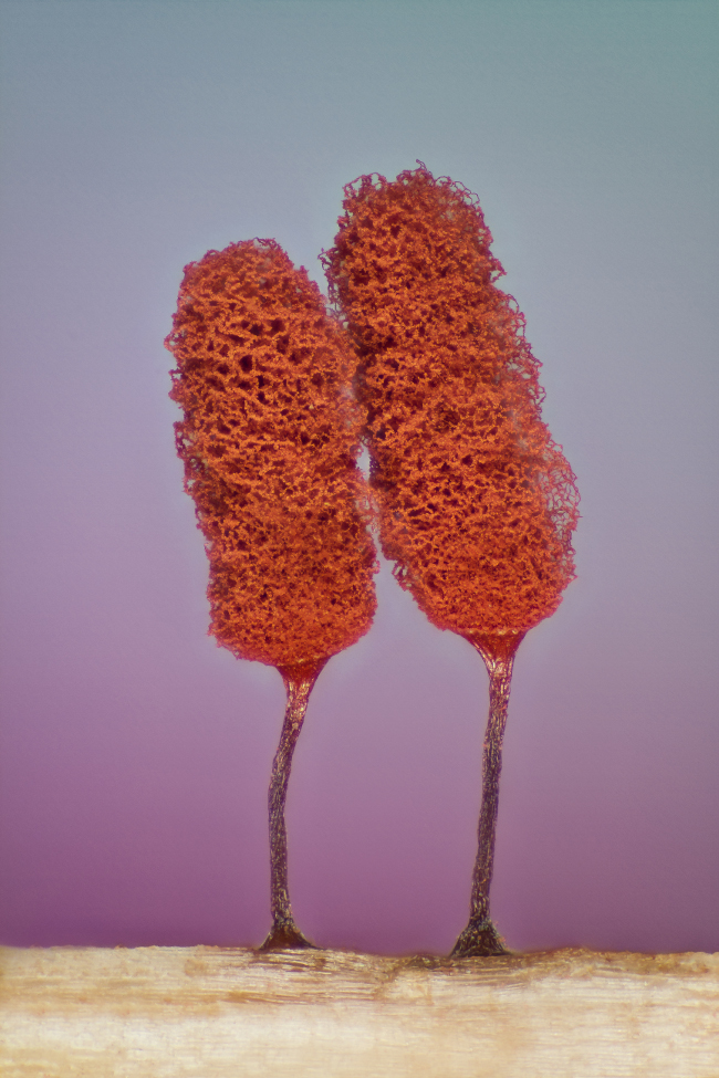
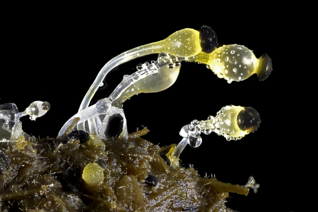
17th place – Slime mould (Mixomicete) in image stacking/reflected light at 5x. Image: Jose Almodovar/Nikon Small World Photomicrography Competition
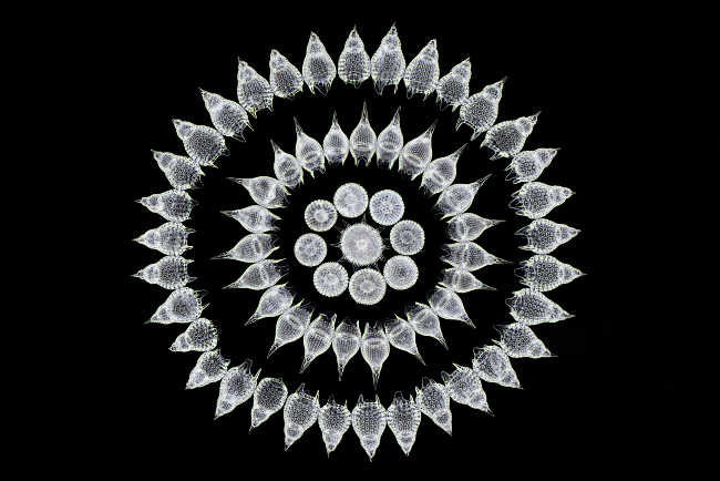
16th place – 65 fossil Radiolarians (zooplankton) carefully arranged by hand in Victorian style in dark field at 100x. Image: Stefano Barone/Nikon Small World Photomicrography Competition
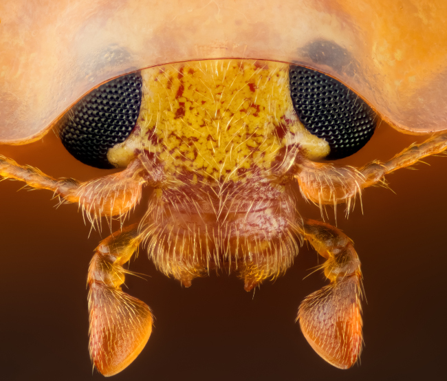
15th place – Head section of an orange ladybird (Halyzia sedecimguttata) in reflected light/focus in stacking at 10x. Image: Geir Drange/Nikon Small World Photomicrography Competition
14th place – Mouse retinal ganglion cells in fluorescence/confocal at 40x. Image: Dr Keunyoung Kim/Nikon Small World Photomicrography Competition
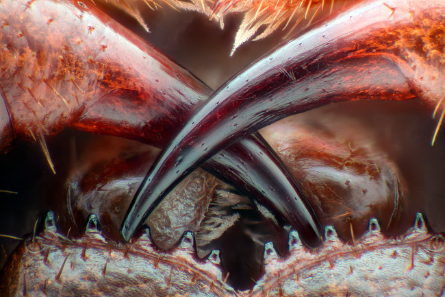
13th place – Poison fangs of a centipede (Lithobius erythrocephalus) in fibre optic illumination/image stacking at 16x. Image: Walter Piorkowski/Nikon Small World Photomicrography Competition

12th place – Human HeLa cell undergoing cell division (cytokinesis). DNA (yellow), myosin II (blue) and actin filaments (red) in structured illumination at 9x. Image: Dr Dylan Burnette/Nikon Small World Photomicrography Competition
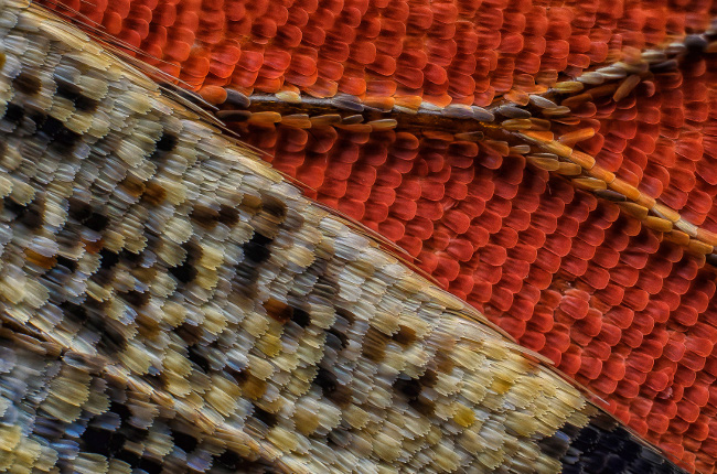
11th place – Scales of a butterfly wing underside (Vanessa atalanta) in macroscopy at 10x. Image: Francis Sneyers/Nikon Small World Photomicrography Competition
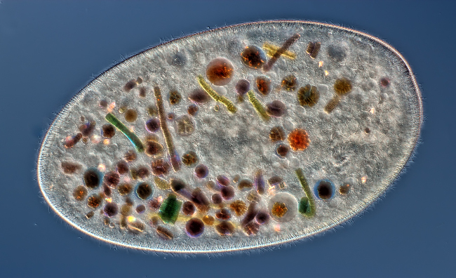
10th place – Frontonia (showing ingested food, cilia, mouth and trichocysts) in differential interference contrast at 200x. Image: Rogelio Moreno Gill/Nikon Small World Photomicrography Competition
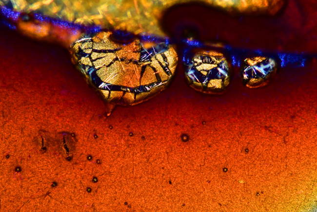
9th place – Espresso coffee crystals in polarised light. Image: Vin Kitayama and Sanae Kitayama/Nikon Small World Photomicrography Competition
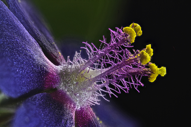
8th place – Wildflower stamens in fibre optic illumination at 40x. Image: Samuel Silberman/Nikon Small World Photomicrography Competition
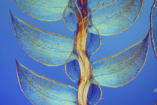
7th place – Leaves of Selaginella (lesser club moss) in differential interference contrast at 40x. Image: Dr David Maitland/Nikon Small World Photomicrography Competition
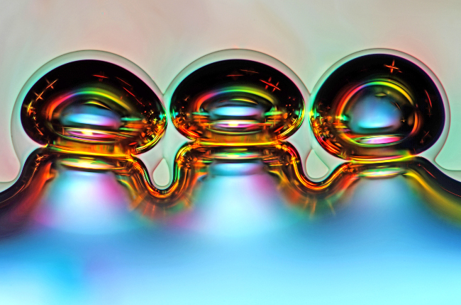
6th place– Air bubbles formed from melted ascorbic acid crystals in polarised light at 50x. Image: Marek Mis/Nikon Small World Photomicrography Competition
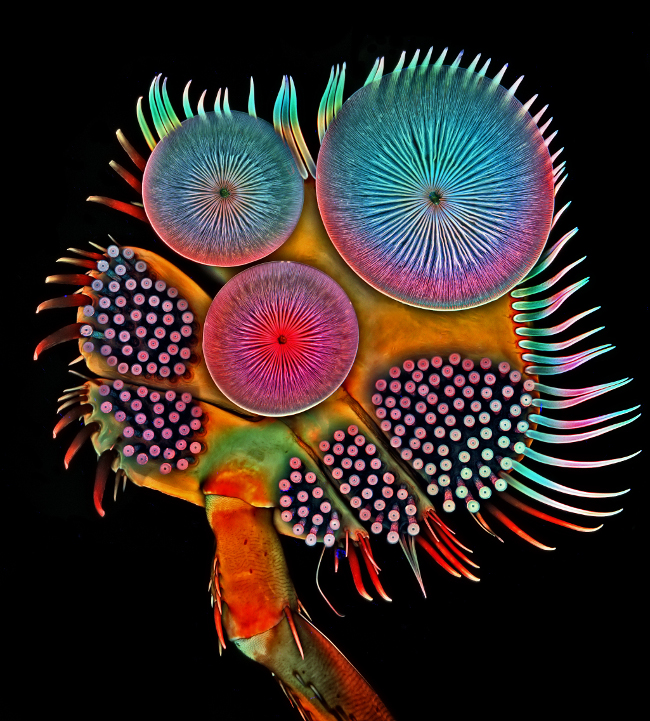
5th place – Front foot (tarsus) of a male diving beetle in confocal at 100x. Image: Dr Igor Siwanowicz/Nikon Small World Photomicrography Competition
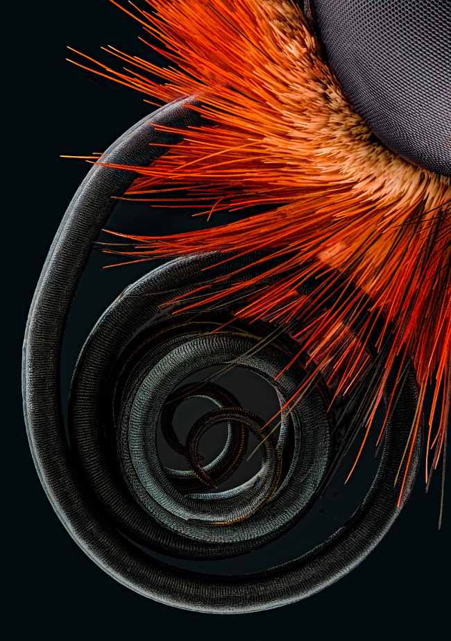
4th place – Butterfly proboscis in image stacking at 6.3x. Image: Jochen Schroeder/Nikon Small World Photomicrography Competition
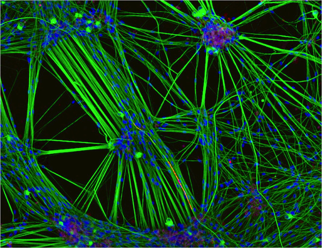
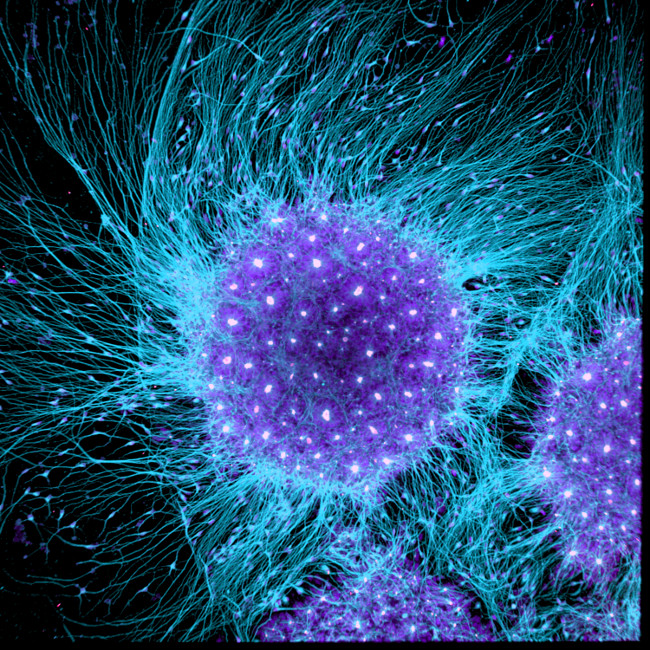
3rd place – Culture of neurons (stained green) derived from human skin cells, and Schwann cells, a second type of brain cell (stained red) in confocal/immunofluorescence/iPSCs at 20x. Image: Rebecca Nutbrown/Nikon Small World Photomicrography Competition
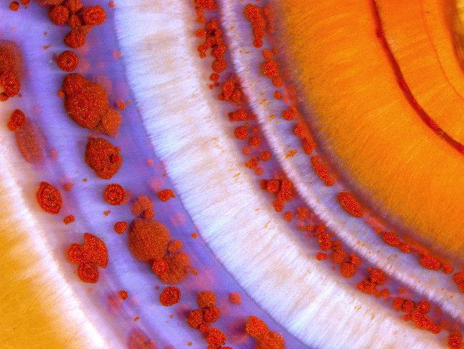
2nd place – Polished slab of Teepee Canyon agate in stereomicroscopy at 90x. Image: Douglas L Moore/Nikon Small World Photomicrography Competition
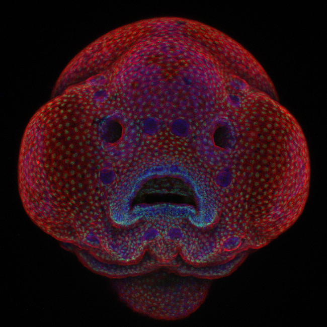
1st place – Four-day-old zebrafish embryo, in confocal at 10x. Image: Dr Oscar Ruiz/Nikon Small World Photomicrography Competition